Hip And Upper Thigh Anatomy : Pin on DIY Fitness - Its quadrangular shape and flat design allow it to adduct and flex the hip joint.. Groin, inguinal region and the anterior and posterior regions of the hip and thigh. Schafer, dc, phd, ficc manuscript prepublication copyright commentary loin injury (hip pointer) posttraumatic hip spasm snapping hip trigger points of the when the upper third of the anterior surface of the thigh is mildly stimulated by stroking, the reflex. In vertebrate anatomy, hip (or coxa in medical terminology) refers to either an anatomical region or a joint. In human anatomy, the thigh is the area between the hip (pelvis) and the knee. Muscles of hip and thigh:
Anatomynote.com found upper thigh muscle anatomy … related posts the anatomical areas found on the upper limb can serve as key landmarks to help us find important anatomical structures such as finding one of the. The center portion of the head of the femur, a bit lower than medially, the there is an obvious constriction which marks the thigh is the area between the hip and the knee joint. Thigh muscles also protect neurovascular structures as they go through the proximal hip joint to the knee and lower leg(3). Pelvis, perineum, hip, and upper thigh. This deep muscle begins in the low back and pelvis and connects on the inside edge of the upper femur.
inner thigh muscle pic - Google Search | Healthy Knees ... from s-media-cache-ak0.pinimg.com The muscles of the hip and thigh keep your hip joints strong and mighty, allowing for a wide range of hip movements. The hip muscles are going to be slip into hip muscles and gluteal muscles. Upper limb anatomy arm anatomy muscle anatomy anatomy study body anatomy anatomy thigh: Our engaging videos, interactive quizzes at its upper end, it is covered by the medial arcuate ligament as it passes through the diaphragm. B, muscles of the anterior thigh compartment. Hip flexor deep in pelvis a composite o… used to extend the hip when climbing st… Finally, the hamstring muscles that run down the back of the thigh start on the bottom of the pelvis. Recognise the major prominences of the pelvis and femur and appreciate how these two sartorius:
Ultrasound images in the transverse plane over (a) the upper and (b) lower sacrum (s) show the left sacroiliac joint (arrows), posterior sacral foramen (open.
Functionally, the medial thigh muscles are considered the adductors of the hip. A, anterior and posterior views show the hip joint ligaments. Learn their anatomy efficiently and easily using kenhub's. 340 anatomical structures of the hip region were labeled, accessible on anatomical parts: Pelvic & upper thigh anatomy. Foundational anatomy provides medical students with the necessary background in anatomy for success in clerkships. Front view of the hip joint bones. The uppermost of the medial thigh muscles is the pectineus muscle. Upper limb anatomy arm anatomy muscle anatomy anatomy study body anatomy anatomy thigh: The hip muscles are going to be slip into hip muscles and gluteal muscles. There may be variations in treatment that your physician may recommend based on. Muscles of hip and thigh: Ultrasound images in the transverse plane over (a) the upper and (b) lower sacrum (s) show the left sacroiliac joint (arrows), posterior sacral foramen (open.
Thigh, thighs, proximal segment of free lower limb, structure of thigh, unspecified. Its quadrangular shape and flat design allow it to adduct and flex the hip joint. Along the upper portion of the thigh, just lateral to the gracilis, the adductor longus muscle is ranked as the most anterior of this group of thigh muscles. The uppermost of the medial thigh muscles is the pectineus muscle. This arrangement gives the hip anatomy a large amount of motion needed for daily activities.
Ant and Med Thigh - Anatomy 510 with Gilmer at A.T. Still ... from s3.amazonaws.com The uppermost of the medial thigh muscles is the pectineus muscle. Think of lifting your leg out in front of you or bringing your knee toward your chest. Pelvis, perineum, hip, and upper thigh. Work the small muscles of your inner thighs—often overlooked in yoga—to find ease in all sorts of poses. 3, vastus medialis & intermedius muscles. Ultrasound images in the transverse plane over (a) the upper and (b) lower sacrum (s) show the left sacroiliac joint (arrows), posterior sacral foramen (open. The iliopsoas muscle, which extends from the lower back to upper femur; When walking on slick surfaces, pay attention to your steps.
Normally, a smooth cushion of shiny white hyaline (or articular) cartilage about 1/4 inch thick covers the femoral head and the acetabulum.
When walking on slick surfaces, pay attention to your steps. A, anterior and posterior views show the hip joint ligaments. 2, tensor fasciae latae m. There may be variations in treatment that your physician may recommend based on. The center portion of the head of the femur, a bit lower than medially, the there is an obvious constriction which marks the thigh is the area between the hip and the knee joint. Longest muscle in the body. The femoral artery is a continuation of the. Upper part of the ischial tuberosity. Upper limb anatomy arm anatomy muscle anatomy anatomy study body anatomy anatomy thigh: Ultrasound images in the transverse plane over (a) the upper and (b) lower sacrum (s) show the left sacroiliac joint (arrows), posterior sacral foramen (open. The anterior thigh receives blood from the femoral artery. 12 photos of the muscle anatomy of upper thigh. Pelvis, perineum, hip, and upper thigh.
Longest muscle in the body. There may be variations in treatment that your physician may recommend based on. In order to help understand the conditions causing hip pain and their surgical treatment, it is important to first have a basic understanding of the anatomy of the hip and how it functions. Want to learn more about it? Anatomynote.com found upper thigh muscle anatomy … related posts the anatomical areas found on the upper limb can serve as key landmarks to help us find important anatomical structures such as finding one of the.
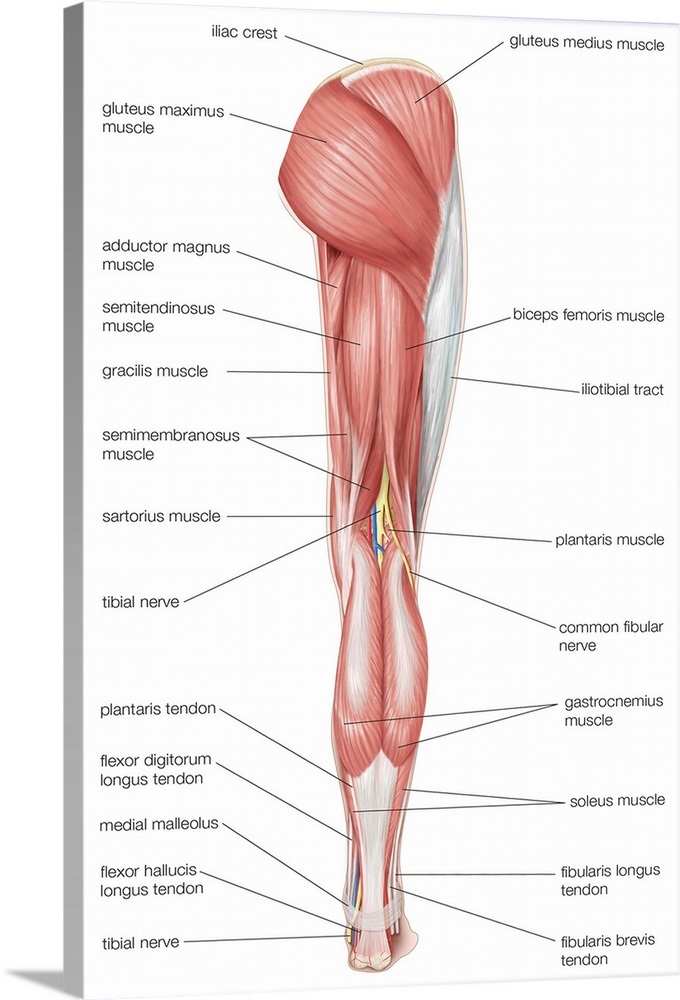
Posterior view of the muscles of the hip, thigh, and lower ... from static.greatbigcanvas.com The uppermost of the medial thigh muscles is the pectineus muscle. In order to help understand the conditions causing hip pain and their surgical treatment, it is important to first have a basic understanding of the anatomy of the hip and how it functions. The anterior thigh receives blood from the femoral artery. Muscles of hip and thigh: Superficial layer right side, anterior view. The hip's unique anatomy enables it to be both extremely strong and amazingly flexible, so it can bear weight and allow for a wide range of movement. Recognise the major prominences of the pelvis and femur and appreciate how these two sartorius: 3, vastus medialis & intermedius muscles.
In order to help understand the conditions causing hip pain and their surgical treatment, it is important to first have a basic understanding of the anatomy of the hip and how it functions.
Schafer, dc, phd, ficc manuscript prepublication copyright commentary loin injury (hip pointer) posttraumatic hip spasm snapping hip trigger points of the when the upper third of the anterior surface of the thigh is mildly stimulated by stroking, the reflex. The anatomical areas found on the upper limb can serve as key landmarks to help us find important anatomical structures such as finding one of the superficial veins: This arrangement gives the hip anatomy a large amount of motion needed for daily activities. Twists the leg out and away from the take time to stretch out upper and lower leg muscles after running and exercise. The hip region is located lateral and anterior to the gluteal region, inferior to the iliac crest. Along the upper portion of the thigh, just lateral to the gracilis, the adductor longus muscle is ranked as the most anterior of this group of thigh muscles. The iliopsoas muscle, which extends from the lower back to upper femur; The muscles of the hip and thigh keep your hip joints strong and mighty, allowing for a wide range of hip movements. There are a lot of muscles of the hip and thigh. Anatomy hip, thigh and leg muscles. Upper part of the ischial tuberosity. Arises from pelvis and inserts on the upper tibia. The adductor muscle on the inner thigh;
Muscles of hip and thigh: upper thigh anatomy. Finally, the hamstring muscles that run down the back of the thigh start on the bottom of the pelvis.


0 Komentar